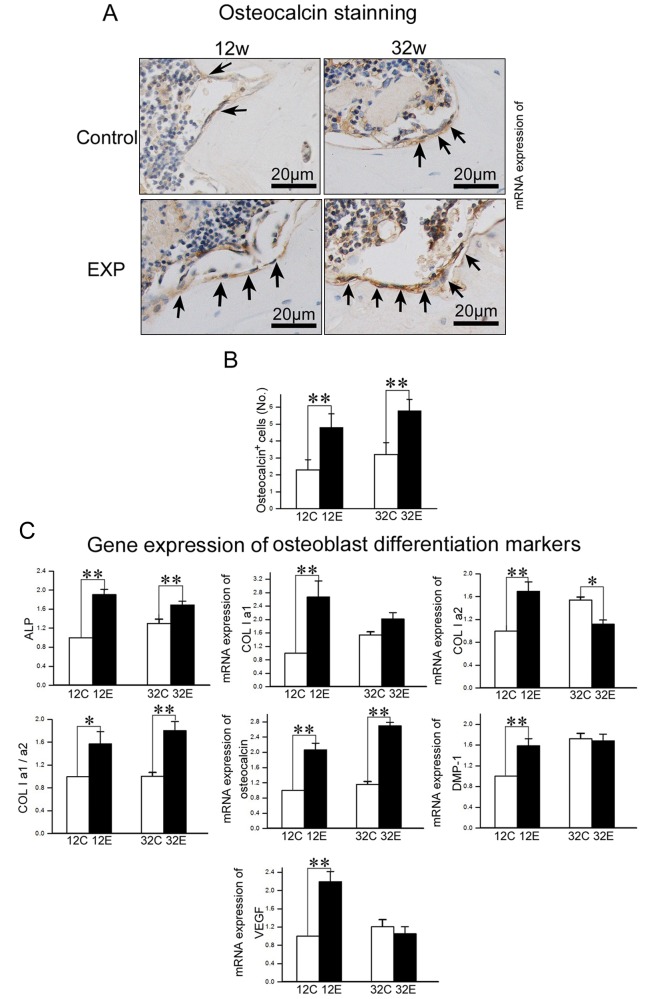
Figure 4.
Analysis of osteoblast activity in TMJ condylar subchondral bone. (A) Osteocalcin immunohistochemical (IHC) staining of TMJ subchondral bone from 12- and 32-week control and experimental groups. (B) The number of osteocalcin-positive cells within the selected area was counted. (C) Real-time PCR analysis of expression of ALP, Col1a1, Col1a2, the ratio of Col1a1/Col1a2, osteocalcin, DMP-1, and VEGF genes from the subchondral bone of the mandibular condylar head in 12- and 32-week control and experimental groups (*p < 0.05, **p < 0.01).

