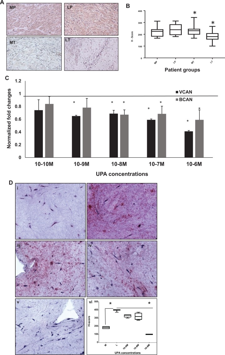
Figure 4.
Ulipristal acetate treatment decreases the expression of proteoglycans in treated leiomyoma (LT) tissue and 3D cultures. A, Primary antibody directed against aggrecan protein is demonstrated in the cytoplasm of positively stained myometrial and leiomyoma patient tissue samples. Both treated myometrium (MT) and LT tissues resulted in a decrease in the expression of aggrecan PG, indicated by less stain intensity for this protein. B, Analysis of the expression of aggrecan (H-scores) in myometrial and leiomyoma patient tissue. Both placebo leiomyoma tissue (255 ± 8.5) and treated myometrial tissue (223.8 ± 8.4) was similar to placebo myometrium (241.9 ± 10.8). Treated leiomyoma patient samples demonstrated a decrease in the expression of aggrecan (172.3 ± 6.7) that was statistically significant (**P < .003). C, Western blot analysis of 3D cultures treated with UPA demonstrate a decrease in both versican and brevican proteoglycans at all UPA concentrations. D, Immunohistochemical analysis of VCAN production in UPA-treated 3D leiomyoma cells stained for versican (i-vi) are shown. (i) Myometrial 3D culture; 0 M, (ii) Leiomyoma culture; 0 M, (iii-v) Leiomyoma treated with UPA at 10−9 M, 10−8 M, and 10−6 M. Note that at UPA concentration of 10−6 M (v) the stain intensity for VCAN is markedly diminished, resembling that of myometrial 3D cultures. (vi) H-score analysis of VCAN images: Myometrium (i; 137.6 ± 29.6), Leiomyoma (ii; 315.4 ± 75.6), 10−9 M (iii; 237.5 ± 58.8), 10−8 M (iv; 321.9 ± 21.5), and 10−6 M (v; 100.0 ± 0) (*P < .03). PG indicates proteoglycans; UPA, ulipristal acetate; VCAN, versican.

