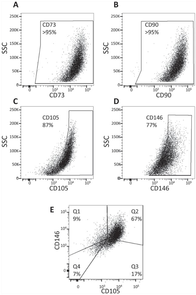
Figure 2.

Scatter plots showing the expression of mesenchymal stem cell markers by multicolor flow cytometry analysis. The majority of cells isolated from intracanal blood following overinstrumentation of the periapical tissues coexpress mesenchymal stem cell markers. After doublet discrimination, CD45-negative cells were analyzed for CD73, CD90, CD105, and CD146 using gates based on the unstained control. CD73 (A) and CD90 (B) were found to be expressed in >95% of cells, whereas CD105 (C) and CD146 (D) were expressed in 87% and 77% of cells, respectively. (E) Of the CD73 and CD90 coexpressing cells, 9% also expressed CD146 but not CD105 (Q1); 67% expressed both CD105 and CD146 (Q2); 17% expressed only CD105 but not CD146 (Q3); and 7% did not express either CD105 or CD146. SSC, side scatter.
