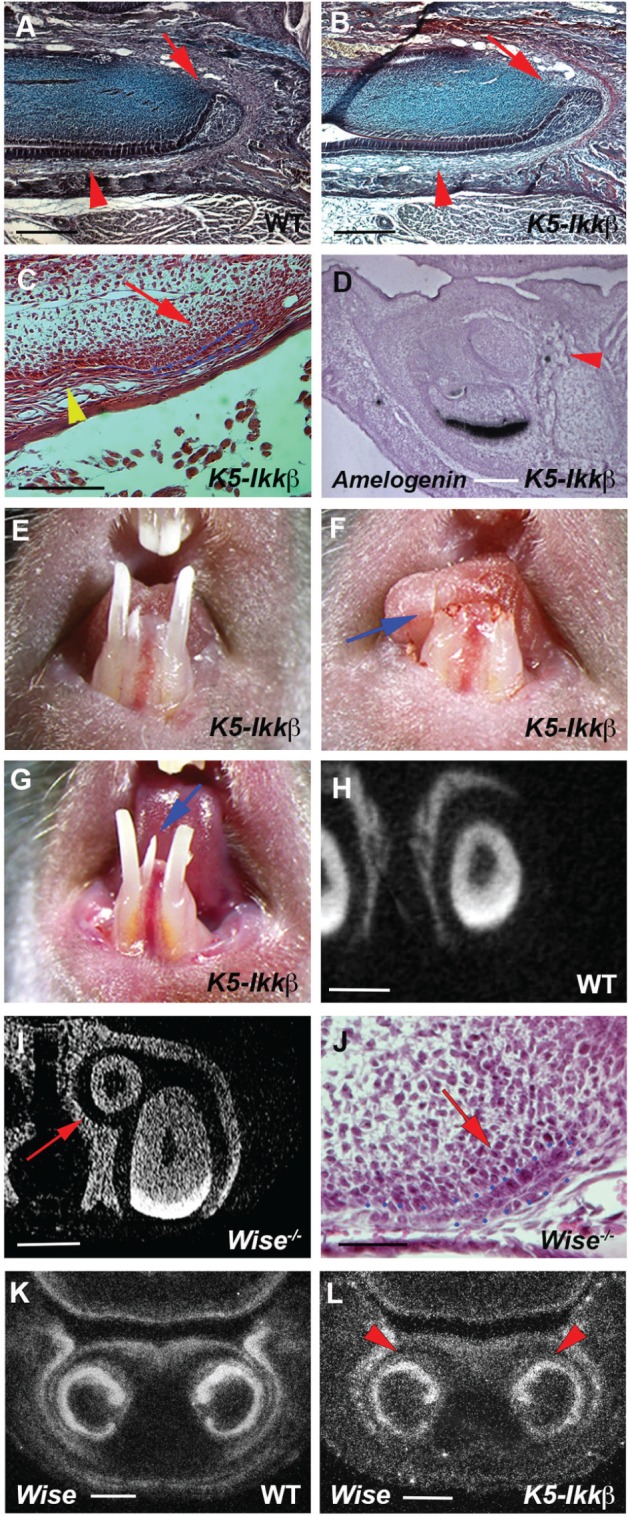
Figure 4.

Lingual incisors in K5-Ikkβ mice. (A–C, J) Sagittal sections showing labial cervical loop in endogenous incisor (arrows in A, B) and lingual incisor (arrows in C, J) of wild-type (A), K5-Ikkβ mice (B, C), and Wise mutant (J) at P5. Arrowheads indicating ameloblasts (A, B) and the lack of ameloblasts (C). (D) Sagittal sections showing Amelogenin mRNA expression in K5-Ikkβ mice. Arrowhead indicating lingual incisor (D). (E–G) Endogenous and lingual incisors before clipping (E), just after clipping (arrow in F), and at 2 wk after clipping (G). Arrow indicating newly formed lingual incisor (G). (H, I) Frontal section based on micro–computed tomography scan in lower incisors of wild-type (H) and Wise mutant (I). Arrow indicates lingual incisor (I). (K, L) Wise expression by radioactive in situ hybridization on frontal sections of wild-type (K) and K5-Ikkβ mice (L) at E15.5. Arrowheads showing Wise expression on the lingual side of endogenous incisor tooth germs (L). The labial cervical loop was outlined by blue dots (C, J). Scale bars: 500 µm (A, B, H, I), 300 µm (C), 150 µm (D, J), and 200 µm (K, L).
