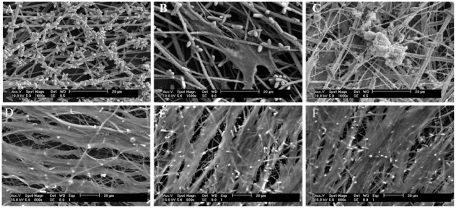
Figure 2.
Scanning electron microscope observation of cell-mediated mineral nodules. (A) Observation of FA-modified PCL nanofiber scaffold. Observation of dental pulp stem cells grown on PCL + FA scaffolds for (B) 7 d and (C) 28 d. At 28 d, densely deposited mineral nodules were observed. Observation of dental pulp stem cells grown on PCL + FA scaffolds for 28 d with specific pathway inhibitors: (D) cyclopamine (the inhibitor of hedgehog), (E) FH535 (the inhibitor of Wnt), and (F) PPP (the inhibitor of insulin). No mineral nodules were seen on the surfaces. FA, fluorapatite; PCL, polycaprolactone.

