-
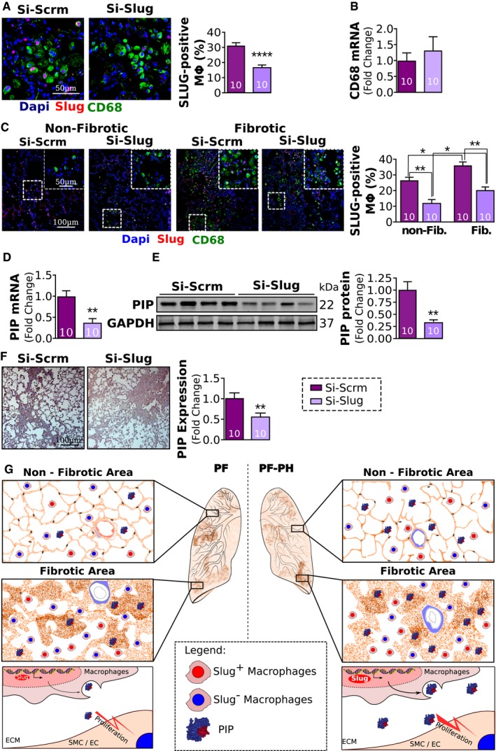
A
Representative images and quantification of Slug‐positive macrophages (Slug, red; CD68 green).
-
B
Relative expression of CD68 mRNA normalized to GAPDH.
-
C
Representative images and quantification of Slug‐positive macrophages between non‐fibrotic and fibrotic areas of the lung.
-
D, E
Relative expression of PIP mRNA (D) and protein (E) normalized to GAPDH.
-
F
Representative images and quantification of PIP immunohistochemistry.
-
G
Proposed mechanism. In pulmonary fibrosis, vascular wall thickening is mainly seen in fibrotic areas of the lung, while in PH secondary to PF, vascular remodeling is seen in both fibrotic and non‐fibrotic areas of the lung. The extension of vascular wall thickening to the non‐fibrotic areas of the lung in PF‐PH is concomitant with an increased expression of Slug and PIP in the lungs in both fibrotic and non‐fibrotic areas. At the cellular level, Slug upregulation in macrophages in PF‐PH leads to increased PIP expression in the extracellular space, which in turn triggers SMC proliferation leading to vascular wall thickening observed in fibrotic and non‐fibrotic areas of the lung (ECM: extracellular matrix; SMC/EC: smooth muscle cells and endothelial cells).
Data information: Values are expressed as mean ± SEM. The number of samples per group for each experiment is included within each bar graph. Statistical tests: panels (A, B, and D–F):
t‐test; panel (C): ANOVA (*
P < 0.05, **
P < 0.01, ****
P < 0.0001).