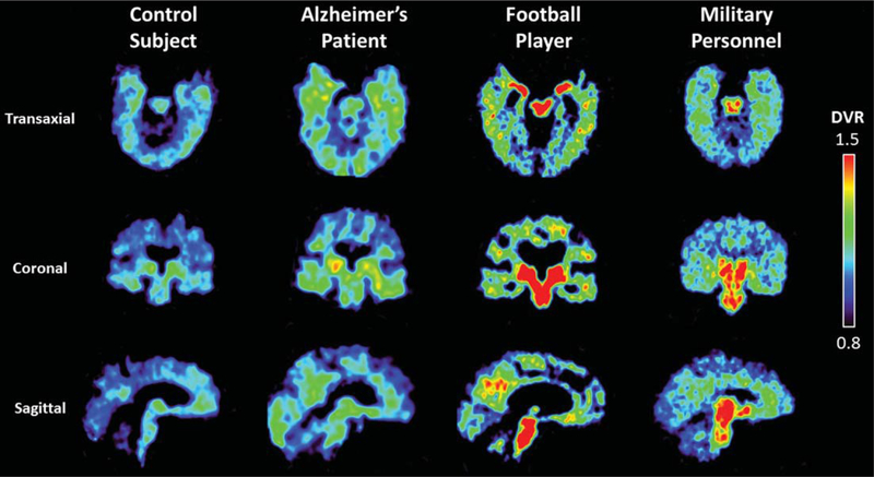
Fig. 1.
Examples of FDDNP-PET DVR transaxial, coronal, and sagittal images of a cognitively healthy individual, Alzheimer’s disease patient, football player, and a military subject. The Alzheimer’s disease patient shows higher DVR signals in parietal, temporal and frontal regions compared to the healthy individual, who may have neuropathology deposition given the age of the subject (80 years) [11]. The football player and military subject show higher amygdala, midbrain, and other subcortical binding compared with the healthy individual and Alzheimer’s disease patient. The healthy individual showed some mild cortical binding typical of other healthy individuals age 70 and older (or darker shades in greyscale) [10]. Warmer colors (or darker shades in greyscale) indicate higher FDDNP binding.

