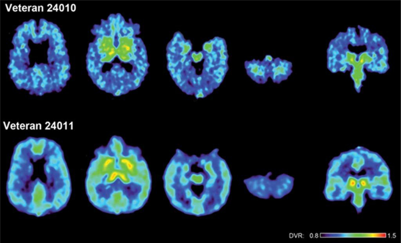
Fig. 2.
FDDNP DVR parametric images of the brains of two war veterans with histories of multiple blast concussions (mTBIs) during their war zone deployment. The upper row shows a 48-year-old man (veteran 24010) and the lower row a 36-year-old man (veteran 24011). Left four images in each row show transaxial brain images from top of the brain to the bottom. The right image shows a coronal cut through the midbrain. (This figure was originally published in Barrio et al., 2015 [16].)

