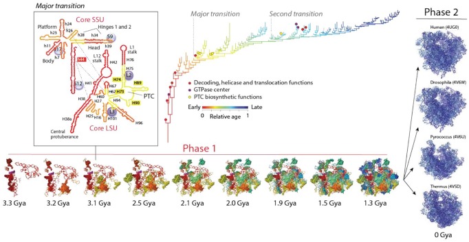
Figure 3.
The biphasic history of the ribosome. An evolutionary timeline of ribosomal RNA (rRNA) and proteins (r-proteins) inferred directly from phylogenomic data shows 2 evolutionary phases. During an initial phase (phase 1), helical structures of rRNA and r-proteins accreted to form a universal ribosomal core. The second phase of ribosomal evolution (phase 2) started 1.3 Gya (or earlier) when the universal core diversified alongside with evolving organismal lineages. The phylogenomic tree describes the accretion of rRNA helical stems and is colored according to relative age. Every new branch reflects the addition of a new part to the whole. Only selected functional taxa are labeled in the tree with colored circles. The first RNA structures to accrete include the head and ratchet, the central protuberance, and stalks, which are involved in ribosomal dynamics. Early structures are also involved in energetics, decoding, helicase activity, and translocation. The peptidyl transferase center (PTC) that is responsible for protein biosynthesis accretes later in time (in yellow), whereas RNA helices gradually gained interaction with r-proteins to form a processivity core 2.8 to 3.1 Gya at a time when a crucial “major transition” in ribosomal evolution brought small and large subunits (SSU and LSU) together through protein structural stabilization, interaction surfaces, and formation of intersubunit bridges. The inset shows secondary structure representations of the primordial ribosomal ensemble, with r-proteins visualized as bubbles and bridge interactions as dashed blue lines. This initial proto-ribosome served as center for coordinated ribonucleoprotein accretion to form a highly processive universal ribosome core during a “second transition” that took place 2.4 Gya. A molecular clock of folds linked structural and geological timescales.

