
Figure 3.
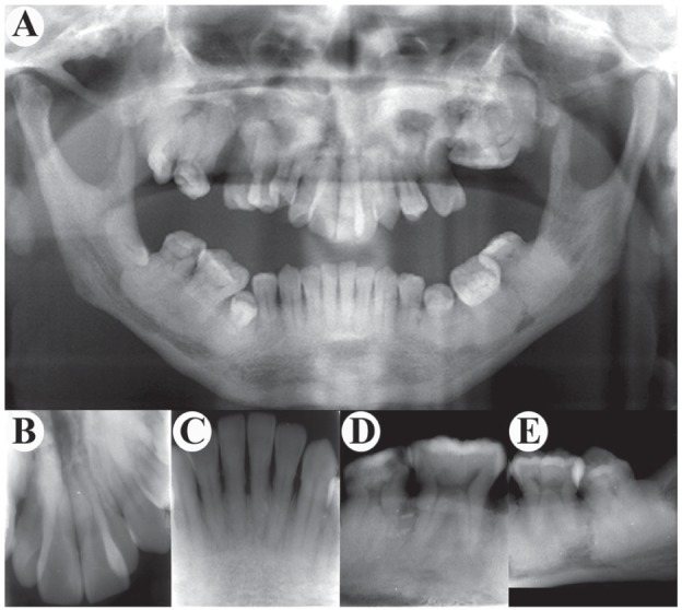
Radiologic findings of teeth. (A) Orthopantomography displays hypoplasia of maxilla and mandible with increased bone density and loss of normal trabeculae. It also shows a dense clump on the left maxillary molar area, obscure nasal cavity, maxillary sinuses, flattened mandibular angle, and the high radiodensity around the roots of lower molars. (B, C) Front teeth show relatively clear root boundaries. (D, E) Mandibular molars display narrowed pulp cavity, stenosis, or atresia in root canal. The most impressive feature is the extensive periradicular high-density clumps whose density appears to be the cortical bone.