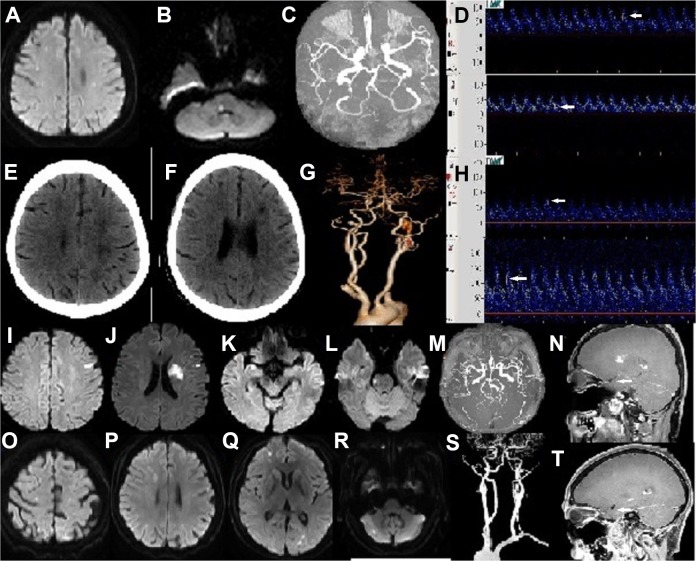
Fig 1.
Patients with the hypercoagulability stroke mechanism (Patients1–4). Patient 1. 42/F/SLE. (A, B) Diffusion-weighted images showed multiple acute infarcts in the bilateral cerebral hemispheres and pons; (C) MR angiography showed normal cerebral arteries; (D) White arrows showed ESs in the LMCA and RMCA. Patient 2. 51/F/SLE. (E, F) Brain CT showed multiple infarcts in bilateral cerebral hemispheres; (G) CT angiography showed normal cerebral arteries; (H) White arrows showed ESs in the LMCA and RMCA. Patient 3. 37/M/SLE. (I, J, K, L) Diffusion-weighted images showed multiple acute infarcts in the bilateral cerebral hemispheres and pons; (M) MR angiography showed normal cerebral arteries; (N) HRMRI (sagittal view) showed normal MCA without circumferential vessel wall thickening or enhancement (white arrow). Patient 4. 58/F/MCTD. (O, P, Q, R) Diffusion-weighted images showed multiple acute infarcts in the bilateral cerebral hemispheres and left cerebellum; (S) CT angiography showed normal cerebral arteries; (T) HRMRI (sagittal view) showed normal MCA without circumferential vessel wall thickening or enhancement (white arrow).
CT: computed tomography; ES: embolic signal; F: female; HRMRI: high-resolution magnetic resonance imaging; LMCA: left middle cerebral artery; M, male; MCTD: mixed connective tissue disease; RMCA: right middle cerebral artery; SLE: systemic lupus erythematosus.

