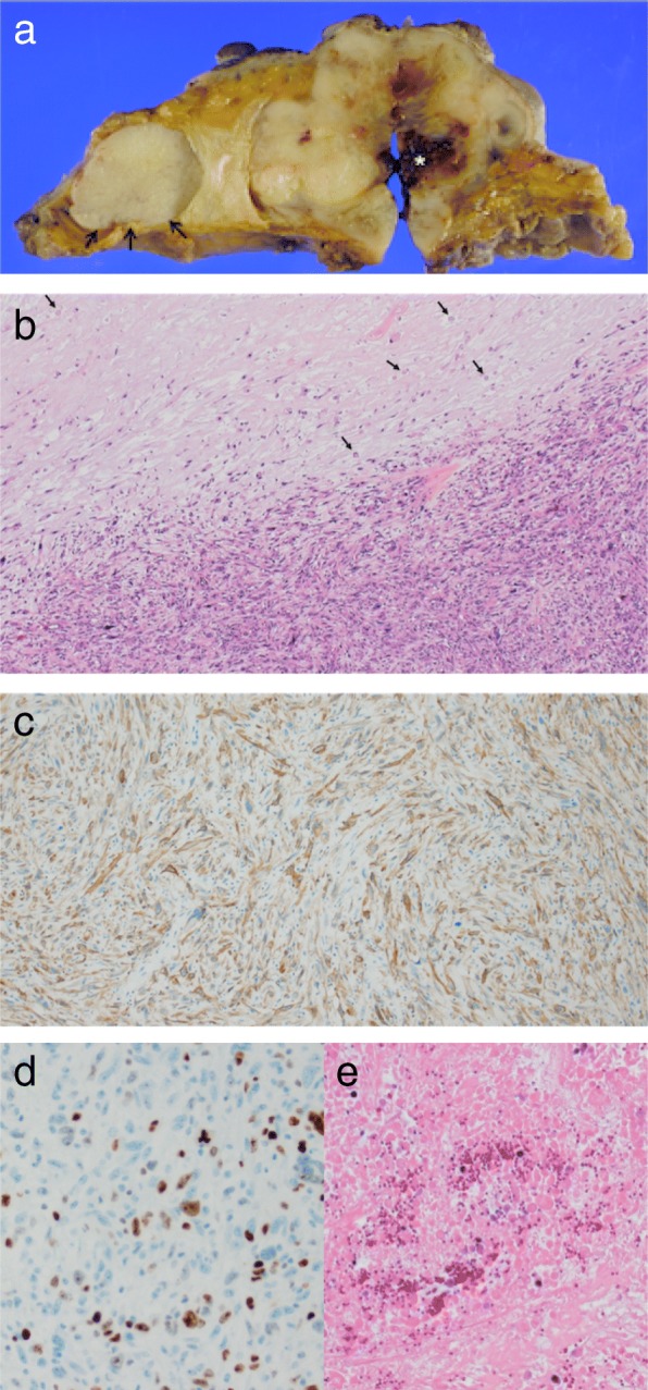
Fig. 2.

a A cross-sectional view of the tumor. Pre-treatment necrosis and hemorrhage is recognized in the center of tumor (asterisk). The mass to the medial side of the main tumor is a benign fibroadenoma (arrow). b On histological examination, nuclear atypia and tight fascicular proliferation of highly cellular spindle tumor cells in the hypercellular areas (high-power view). It also shows the necrotic fibrosis and the weakly stained nucleus known as ghost cells caused by neoadjuvant chemotherapy in hypocellular areas (arrow). c The neoplastic cells are reactive for alpha smooth muscle actin on immunohistochemistry. d The Ki-67 labelling index was approximately 20%. e Necrotic tissue is replaced by granulation and fibrous connective tissue (high-power view)
