
Fig. 3.
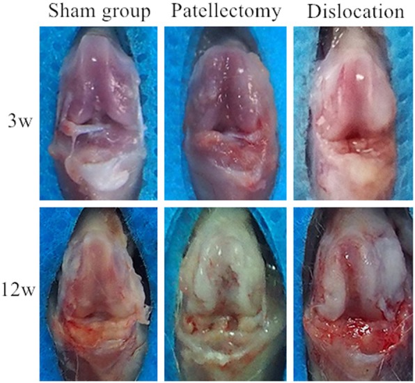
Morphology of cartilage on the trochlear groove. Normal articular cartilage of the trochlea was shown at 3 weeks in the sham group. The shape of the trochlear cartilage was blunt at 3 weeks in the PG. The cartilage on the lateral trochlear facets was thicker than the center and the medial at 3 weeks in the PG. The articular cartilage degenerated at 12 weeks in the PG. After 12-week abnormal patellar tracking by the surgical-induced dislocation, the cartilage in the center of trochlea had shown apparent degeneration and apparent proliferative tissue on the lateral trochlear facets