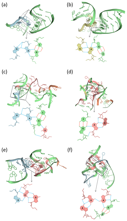
Figure 6.
Molecular images of multiplets with two or more modes of G·A pairing in the complex of tetracycline with the U1052G-mutated 70S Escherichia coli ribosome.69 Loops incorporating m−M sheared pairs are linked by different associations of G and A (highlighted within boxes) to other secondary structural units. A local representation of the linked bases is shown below each global depiction of associated secondary structures. Examples include: (a) a tetraplex with m+m pairing between a hairpin loop and a double-helical stem; (b) a tetraplex with m+m pairing between an internal loop and a double-helical stem; (c) a pentaplex with m+W pairing between a hairpin loop and a double-helical stem that is linked in turn to a junction; (d) a tetraplex with m−m link pairing between an internal loop and a 5-way junction; (e) a triplex with m−W pairing between a hairpin loop and a 3-way junction; (f) a pentaplex with .–M and m−W pairing within a 7-way junction. G·A pairs and secondary structural motifs color-coded as in Figure 2. See Table S3 for respective Protein Data Bank identifiers, chain names, and residue numbers of depicted G·A-linked multiplets. Color-coding of bases and hydrogen bonds matches that in the corresponding secondary structural diagrams in Figure S8.

