
Figure 2.
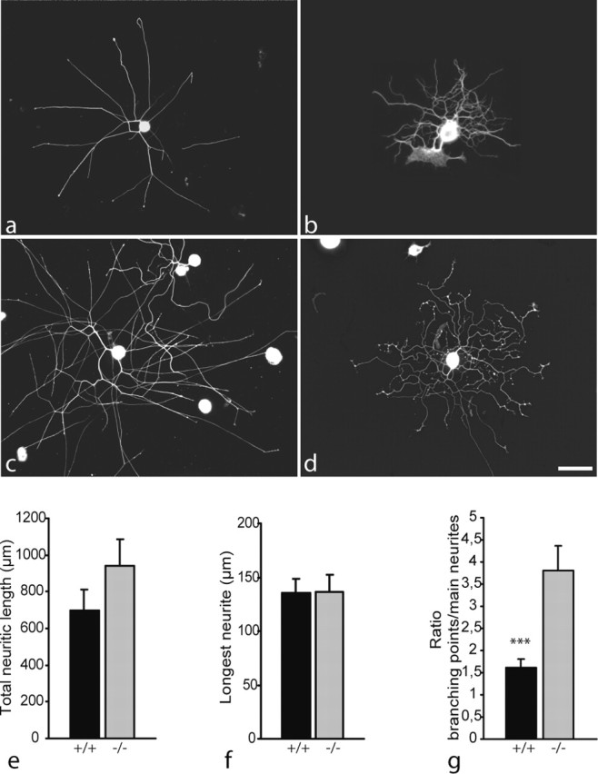
Morphology of neurites regenerating from dissociated wild-type (a, c) and map1b-/- (b, d) DRG neurons after 12 hr (a, b) and 24 hr (c, d) in vitro, visualized by tubulin immunostaining (Tuj-1 antibody). As observed in DRG explant cultures, neurites lacking MAP1B appear more curled than wild-type neurites. Note also the high number of branching points present on map1b-/- neurons. Scale bar, 100 μm. e-g, Quantitative analysis after 12 hr in vitro of total length of neuritic trees (e) and length of the longest neurite (f). No significant difference was observed between cultures of map1b-/- and map1b+/+ neurons. g, In contrast, the number of branching points per main neurite is significantly higher for map1b-/- than for map1b+/+ neurons (***p < 0.001; ANOVA-1).