Figure 6.
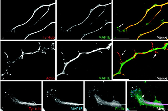
MAP1B immunolocalization in growth cones and at branching points. a, Double immunostaining for tyrosinated MTs (Tyr-tub; red) and MAP1B (green) shows that both proteins colocalize along the axon shaft and in the growth cone, in which individual MT bundles extend into the P-domain (arrow). Note the splayed aspect of both cytoskeletal components in the distal region and at branching points. b, Double immunostaining with anti-actin (red) and MAP1B (green) antibodies reveals colocalization of both proteins along the neurites in filopodia and newly formed branches (arrows). c, Triple immunostaining for tyr-tubulin (red), MAP1B (blue), and actin-phalloidin (green), demonstrating the colocalization of MAP1B with tyrosinated MTs (pink in merge panel; arrow) in the growth cone and with AFs (light blue; arrowhead) in the P-domain. Scale bar, 10 μm.

