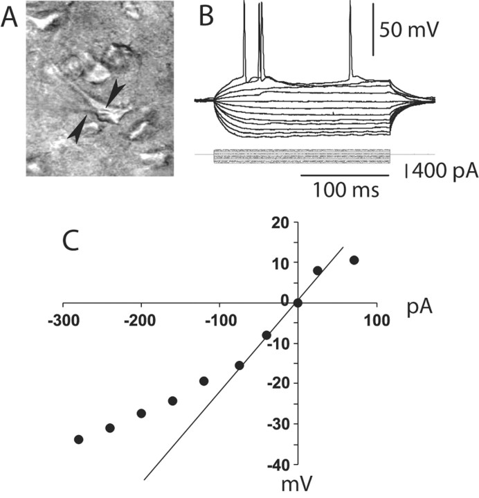
Figure 1.
Whole-cell recording from PFC pyramidal neurons. A, IR-DIC image of a layer V medial PFC pyramidal neuron recorded from a 300-μm-thick PFC slice (P52). The arrows point to the shadow of the recording electrode. B, Typical response to depolarizing and hyperpolarizing somatic current pulses (200 msec; from –300 to +100 pA). C, Current–voltage plot from the traces shown in B. Currents larger than –100 pA yielded a marked inward rectification.

