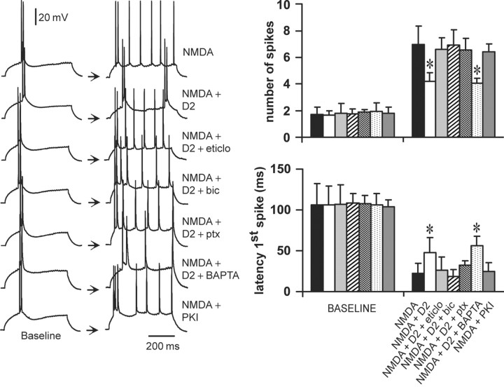
Figure 7.
D2–NMDA interactions involve GABAA receptors. Left, Traces of responses to current injection illustrating changes in action potential firing observed in the different experimental conditions. From top to bottom, the traces are representative recordings obtained with NMDA alone (4 μm), NMDA plus quinpirole (0.4 μm), NMDA plus quinpirole plus the D2 antagonist eticlopride (20 μm; eticlo), NMDA plus quinpirole plus the GABAA antagonist bicuculline (10 μm; bic), NMDA plus quinpirole plus the GABAA antagonist picrotoxin (10 μm; ptx), NMDA plus quinpirole plus BAPTA-containing electrode (2 mm), and NMDA plus PKI [5–24]-containing electrode (20 mm). Right, Bars graphs summarizing the changes in number of evoked spikes and first spike latency in each experimental condition. The inhibitory effect of quinpirole on NMDA responses was blocked by eticlopride (20 μm; n = 5), by bicuculline (10 μm; n = 6), and by picrotoxin (10 μm; n = 6) but not in recordings with electrodes containing the calcium chelator BAPTA (n = 6). Intracellular application of the PKA inhibitor PKI-[5–24] (n = 5) failed to mimic the effects of quinpirole. *p < 0.001; Tukey post hoc test after significant ANOVA.

