
Figure 3.
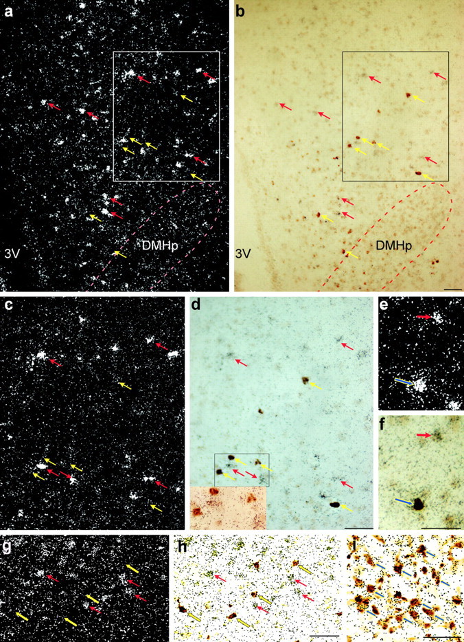
Expression of MC4Rs, GAD67, and NPY mRNA in the rat brain. a, b, Low-power dark-field (a) and bright-field (b) photomicrographs of DMH showing the distribution of MC4Rs (silver grain clusters, red arrows) and NPY neurons (brown cytoplasmic deposit, yellow arrows) scattering next to each other around the compact zone of DMH (DMHp). No apparent colocalization between the two signals was observed in this area. 3V, Third ventricle. c, d, High-power photomicrographs of the boxed areas in a and b to further illustrate that no colocalization of MC4R and NPY was found in the DMH. The inset in d depicts the high-magnification bright-field photomicrograph of the area delineated by the black rectangle in d. e and f show an example of MC4R- and NPY-coexpressing neurons (indicated by the blue arrows) identified in the cerebral cortex. g, h, Low-power dark-field (g) and bright-field (h) photomicrographs of DMH showing GAD67 mRNA-positive neurons (brown cytoplasmic deposit, yellow arrows). Note that little colocalization was observed between the two signals in the DMH. i, Low-power brightfield photomicrograph of the ARH showing that the majority of NPY neurons (brown cytoplasmic deposit, indicated by the blue arrows) coexpressed GAD67 (clusters of black speckles, indicated by the blue arrows). Scale bars, 100 μm.