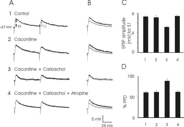
Figure 7.
EPSPs evoked in PH neurons by paired pulses presented to the PPRF and their presynaptic modulation by carbachol and other cholinergic drugs. A, Sample of EPSP records evoked by paired stimuli (S1–S2; interval, 70 msec) presented to the ipsilateral PPRF in the control condition (row 1) and during caconitine (row 2; 0.1 μm), caconitine plus carbachol (row 3; 25 μm), and caconitine plus carbachol plus atropine (row 4; 1.5 μm). Note that carbachol depressed S1- and S2-evoked EPSPs and that the effect was reversed by atropine. Membrane potential of the postsynaptic PH neuron was maintained at its resting level by current injection to cancel out the postsynaptic depolarization evoked by carbachol. B, Superimposition of EPSPs obtained by S1 (black recordings) and S2 (gray recordings) stimuli shown in A. C, Histograms with mean values for EPSP amplitude (in millivolts; n = 10) obtained for S1 presentations. D, Paired-pulse depression (PPD) test. Histograms with mean values (in millivolts; n = 10) obtained for the equation %PPD = S2/S1 × 100, indicating a significant recovery, evoked by carbachol, of the depression induced by the second stimulus (S2). These results suggested a presynaptic action of carbachol.

