
Figure 8.
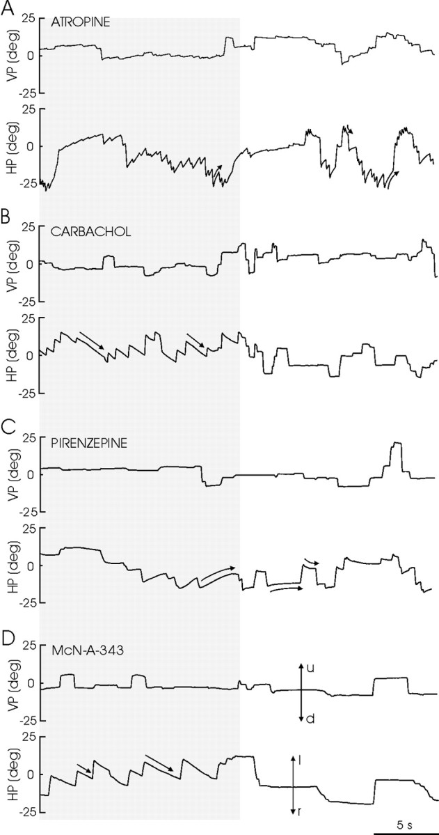
Recordings of left eye position in the vertical (VP) and horizontal (HP) planes obtained in the alert cat in complete darkness (gray lane) and in light, after the injection in the left PH of the indicated drugs. Doses and times after injection were: carbachol (B): 1.5 pmol, 12.8 min; McN-A-343 (D): 20 pmol, 4.5 min; atropine (A): 8 mmol, 20.3 min; pirenzepine (C): 0.12 pmol, 4.9 min. Eye position is plotted as degrees of rotation in the horizontal plane, positive to the left (l) and up (u) and negative to the right (r) and down (d). The zero (0) indicates the central position of the eye in the orbit. The curved and straight arrows indicate the characteristics of eye movement induced by the drugs.