
Figure 3.
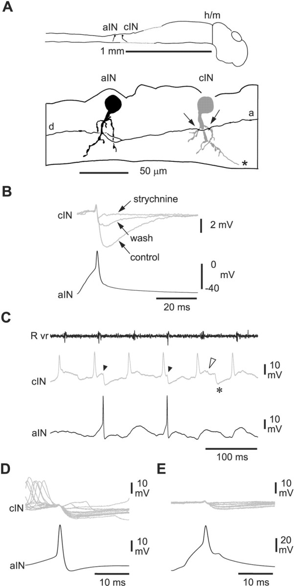
Inhibition from aINs is glycinergic and can produce IPSPs in CPG neurons during swimming. Recordings from an aIN and a cIN. A, Tracings to show the anatomy of an aIN (black) and a cIN (gray) with possible synaptic contacts (arrows) from the ascending (a) axon of the aIN. The aIN also has a descending axon branch (d). The cIN axon is not shown beyond the asterisk, where it crosses the cord ventrally. B, Impulses evoked in the aIN by current injection produce IPSPs in the cIN that are blocked by 5 μm strychnine, with partial recovery on wash. Each trace is the average of seven traces. C, Six cycles of swimming initiated by a current pulse to the skin and monitored from a ventral root on the right side (Rvr). The cIN fires a single impulse and receives IPSPs on most swimming cycles, revealed by injection of depolarizing current (asterisk, mid-cycle IPSP). The aIN fires less reliably. Two early-cycle IPSPs (arrowheads) in the cIN follow spikes in the aIN; another early-cycle IPSP does not (open arrowhead). D, Synchronizing records to the aIN spikes shows short latency IPSPs in the cIN follow most aIN spikes. Note that these IPSPs and the aIN spikes are not similarly time-locked to the preceding cIN spikes. E, IPSPs in the cIN follow spikes evoked in the aIN by current injection at the same latency as those during swimming in D. The IPSPs are smaller than in D because the cIN is less depolarized.