
Figure 1.
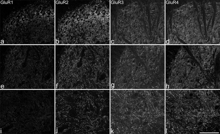
Immunostaining for GluR1-4 in the spinal cord after pepsin treatment. a-d show the medial part of the superficial dorsal horn (corresponding to laminas I-III). e-h include the medial part of laminas IV and V. i-l are from lamina IX. GluR1-immunoreactive puncta are numerous in lamina II, present at lower density in other parts of the dorsal horn, and are virtually absent in lamina IX. GluR2-immunoreactive puncta are present in all parts of the dorsal horn, but the labeling is strongest in lamina II. Punctate labeling with both GluR3 and GluR4 antibodies is seen throughout the gray matter but is least dense in the superficial dorsal horn. Images of GluR1 and GluR2 are taken from one section, and those for GluR3 and GluR4 are taken from another. Each image was obtained from a projection of three optical sections at 0.5 μm z-separation. Scale bar, 100 μm.