
Figure 3.
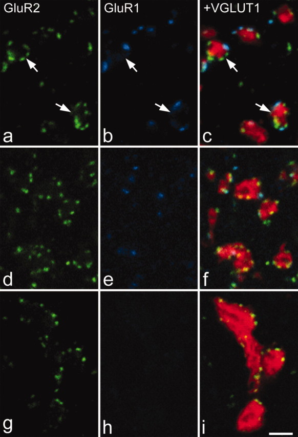
GluR1 and GluR2 puncta associated with VGLUT1-immunoreactive boutons. In each case, GluR2 immunostaining is shown in green in the left panel, GluR1 in blue in the center panel, and these have been merged with VGLUT1 staining (red) in the right panel. a-c are from lamina IIi. d-f are from lamina IV. g-i are from lamina IX. In all cases, the VGLUT1 boutons contact GluR2-immunoreactive puncta. In the dorsal horn, some of these puncta are also GluR1 immunoreactive. In lamina IIi, these puncta often surround the VGLUT1 bouton, with an appearance that is suggestive of a glomerular arrangement (arrows). Note that occasional overlap between GluR-immunoreactive puncta and axonal staining is likely to be attributable to either invagination of a dendrite into a bouton or obliquity of the synapse with respect to the plane of section. All images were obtained from single optical sections. Scale bar, 2 μm.