
Figure 4.
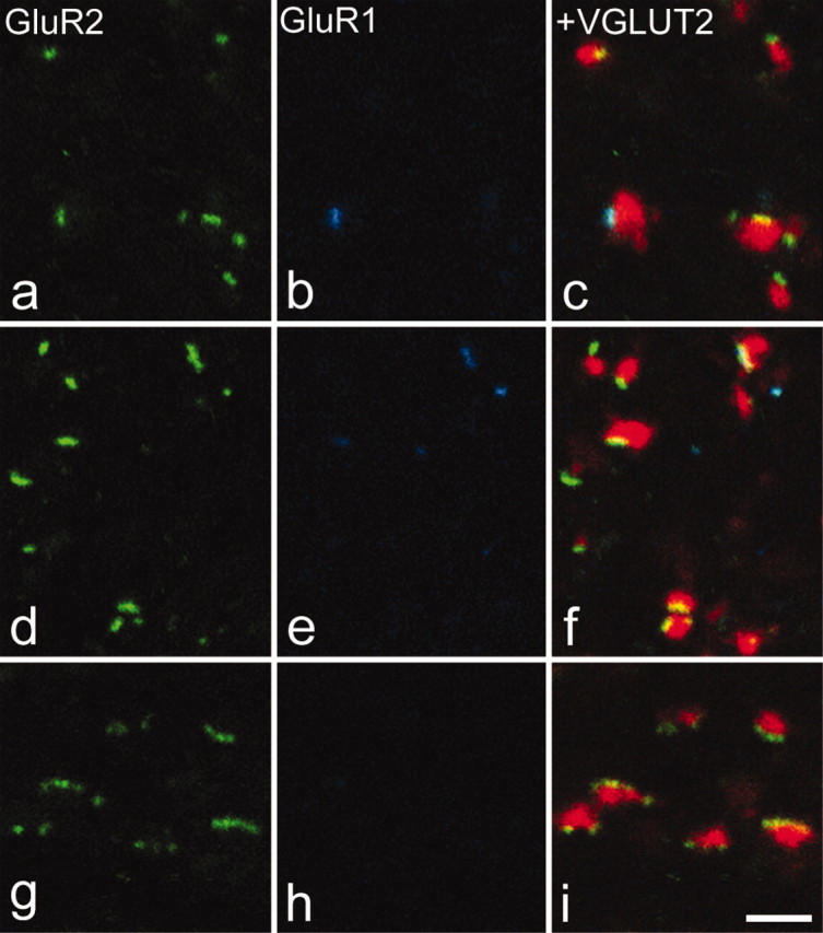
GluR1 and GluR2 puncta associated with VGLUT2-immunoreactive boutons. In each case, GluR2 immunostaining is shown in green in the left panel, GluR1 in blue in the center panel, and these have been merged with VGLUT2 staining (red) in the right panel. a-c are from lamina I. d-f are from lamina V. g-i are from lamina IX. Note that VGLUT2 boutons are associated with few GluR2-immunoreactive puncta and that some of the puncta in contact with these boutons in the dorsal horn are also GluR1 immunoreactive. All images were obtained from single optical sections. Scale bar, 2 μm.