
Figure 7.
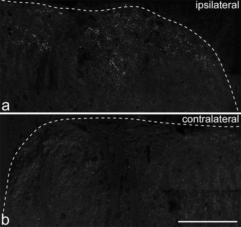
Immunostaining for GluR1-pS845 in a section from L4 in a rat that received an intraplantar capsaicin injection. a shows the medial part of the superficial dorsal horn on the left side (ipsilateral to the injection), and b shows the corresponding part of the right dorsal horn. The dotted line indicates the border between gray and white matter. Numerous immunoreactive puncta are visible in the superficial laminas on the ipsilateral side, but these are rarely seen on the contralateral side. Each image was obtained from a projection of 11 optical sections at 0.5 μm z-separation. Scale bar, 100 μm.