
Figure 3.
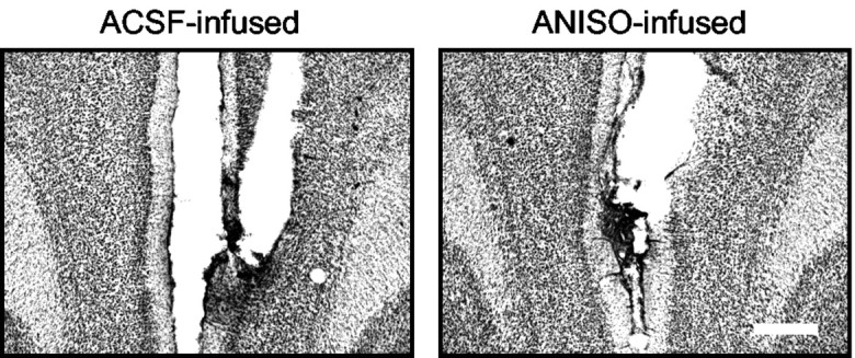
ANISO did not lesion the mPFC. Representative photomicrographs show mPFC tissue from rats infused with ACSF (left) or ANISO (right). There was no evidence of increased cell loss or gliosis in the ANISO animals. Scale bar, 0.5 mm.
Official websites use .gov
A
.gov website belongs to an official
government organization in the United States.
Secure .gov websites use HTTPS
A lock (
) or https:// means you've safely
connected to the .gov website. Share sensitive
information only on official, secure websites.
ANISO did not lesion the mPFC. Representative photomicrographs show mPFC tissue from rats infused with ACSF (left) or ANISO (right). There was no evidence of increased cell loss or gliosis in the ANISO animals. Scale bar, 0.5 mm.