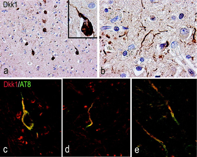
Figure 7.

Association of DKK1 expression and hyperphosphorylated tau in AD temporal cortex. a, b, Neurons with NFT morphology (a) and dystrophic neurites (b) stain for DKK1. c, d, The AT8 antibody reveals the location of hyperphosphorylated tau (green) in neurons (c) and dystrophic neurites (d) expressing DKK1 (red). e, AT8 stains also DKK1-positive white matter fibers.
