
Figure 3.
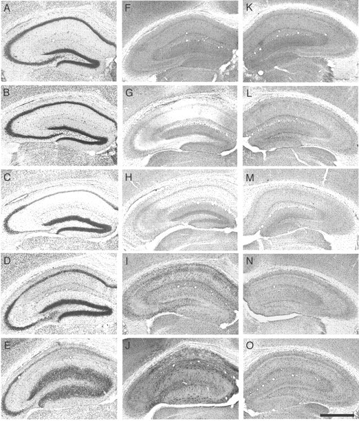
Changes in hippocampal cytoarchitecture ADK immunoreactivity during the course of epileptogenesis. Brains from KA-treated mice were taken at different time points after either intrahippocampal KA or saline injection (n = 4 animals for each time point). Transverse brain sections of the KA-injected brain hemisphere were stained with either cresyl violet (A-E) or for ADK immunoreactivity (F-O). F-J, Ipsilateral side; K-O, Contralateral side. A, F, K, Two hours after saline injection. B, G, L, Two hours after KA injection. C, H, M, One day after KA injection. D, I, N, One week after KA injection. E, J, O, Four weeks after KA injection. Note the biphasic regulation of ADK immunoreactivity after intrahippocampal KA injection (F-J). An initial loss of ADK immunoreactivity within 2 hr after KA injection (G) is gradually followed by a significant overexpression (J), which becomes also evident, although to a lesser degree, within the contralateral hippocampus (O). Scale bar: (in O), A-O, 1000 μm.