Figure 6.
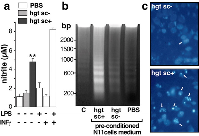
Microglia activation and neurotoxicity induced by PrPsc preconditioned microglia medium. a, Confluent N11 microglial cells were incubated with 5 μl of PBS (white bars), hgtsc- (gray bar), or hgtsc+ (black bar) for 12 hr. Then, nitrite concentration was measured using the Griess reagent and compared with the standard curve. Each histogram is the mean of triplicate determinations from six independent experiments ± SD. b, Representative gel analysis of 10 μg of neuronal DNA from untreated (C) or neurons treated with preconditioned microglia medium as described above. Sizes of the markers are expressed in base pairs (bp) on the left. C, DAPI staining of 4-d-old neurons treated with hgtsc- or hgtsc+ preconditioned microglia culture medium. Phase-contrast micrographs of representative microscopic fields are shown. Magnification, 40×. Arrows indicate apoptotic nuclei.

