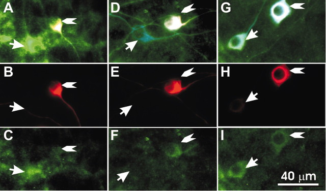
Figure 6.
Basal and apical type II neurons show contrasting α-Kv1.1 staining patterns when compared with neighboring type I spiral ganglion neurons. A-C, Spiral ganglion neurons isolated from the basal cochlea were stained with α-β-tubulin (blue), α-peripherin (red), and α-Kv1.1 (green). A, Superimposed images of the three antibodies. The arrow indicates a type I neuron, and the arrowhead indicates a type II neuron in this and subsequent panels. B, Fluorescent image of α-peripherin staining. C, Fluorescent image of α-Kv1.1 staining. D-I, Two different staining patterns were observed in neurons isolated from the apical region of the cochlea. D-F, An apical type I neuron showed less staining than its neighboring apical type II neuron. D, Superimposed images of the three antibodies. E, Fluorescent image of α-peripherin staining. F, Fluorescent image of α-Kv1.1 staining. G-I, An apical type I neuron showed more staining than the neighboring apical type II neuron. G, Superimposed images of the three antibodies. H, Fluorescent image of α-peripherin staining. I, Fluorescent image of α-Kv1.1 staining.

