Figure 3.

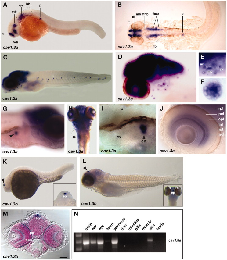
cav1.3a and cav1.3b mRNA expression patterns in the developing and adult zebrafish. mRNA (blue) was detected at embryonic (A, B, 30 hpf; K, 24 hpf) and larval (C-J, L, M, 5 dpf) stages by whole-mount in situ hybridization with cav1.3a (A-J) and cav1.3b (K-M) antisense riboprobes. A, C-G, I-L, Lateral views with anterior to the left and to the right, respectively. B, Dorsal view (anterior to the right) of the same embryo as in A; the embryo was flat mounted. H, Ventral view, anterior to the top. M, Frontal view of a larva cross-sectioned at the level of the epiphysis. E, F, I, J, Close-ups of the following organs (5 dpf): E, ear (focus is on the three cristae; white asterisks denote anterior LL NMs that are not in focus); F, anterior LL NM; I, bean-shaped pancreas. A black asterisk marks expression in a neighboring NM; J, eye. G, H, Arrows mark expression in the pancreas. K, L, Arrows mark expression in the epiphysis organ. K, L, Insets, Anterior dorsal views of the same specimens. t, Telencephalon; vdi, ventral diencephalon; mb, midbrain; ov, otic vesicle; hb, hindbrain; p, pancreas; di, diencephalon; mhb, midbrain-hindbrain boundary; hcp, HC primordium; ac, anterior crista; mc, medial crista, pc, posterior crista; ex, exocrine; en, endocrine; rpl, retinal pigmented epithelial layer; pcl, photoreceptor cell layer; ipl, inner plexiform layer; opl, outer plexiform layer; inl, inner nuclear layer; gcl, ganglion cell layer; ep, epiphysis; lh, left habenula; rh, right habenula. Scale bar: (in M) A, B, 100 μm; C, H, 150 μm; D, 80 μm; E, 65 μm; F, 5 μm; G, 60 μm; I, J, 20 μm; K, 125 μm; L, 200 μm; M, 90 μm. N, A cav1.3a-specific band is detected by RT-PCRs of total RNA from adult brain, ear, eye, pancreas, heart (low levels), and testis tissue homogenates.
