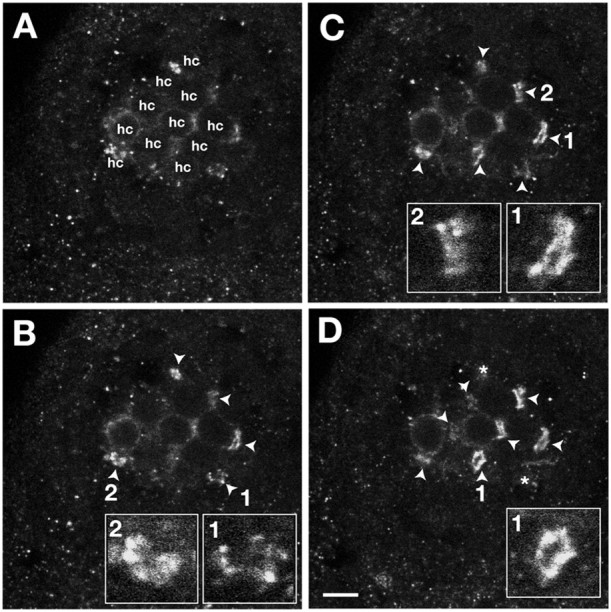
Figure 8.

Number and fine structure of Gem25.2-immunoreactive rings in basal membranes of NM HCs. A-D, Ventral views of four serial confocal sections (0.3 μm thick) through an anterodorsal NM of a 5 dpf wild-type zebrafish larva labeled with Gem25.2. The apical-most section (A) coincides with the mediolateral membrane of the HCs of the NMs; Gem25.2-immunoreactive rings are not yet in focus. The basal-most section(D) is through the basal-most surface of the NM; deeper sections do not contain any additional Gem25.2 rings. The number of HCs was counted in focal planes in which the cell outlines were clearly visible (denoted hc in A). Focal spots in focus are marked by arrowheads and then by asterisks as they leave the focal plane (to mark their former position). In this experiment, we counted 11 HCs and nine focal spots. Insets in B-D are close-ups of individual focal spots. Scale bar, 3 μm.
