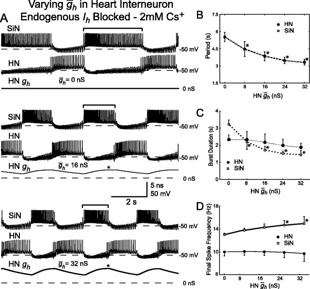
Figure 8.
Variation of ḡh in the heart interneuron (HN ḡh) of a hybrid half-center oscillator with endogenous Ih blocked by 2 mm Cs+. A, Typical activity at three different values of HN ḡh. Voltage traces for the silicon neuron (SiN) and the heart interneuron (HN), and h-current conductance (HN gh) for the heart interneuron are shown. As HN ḡh increases, the burst duration (indicated by brackets) of the unaltered silicon neuron decreases. The asterisks indicate when during the inhibited phase h-current first reaches its maximal level of activation. B, Increasing HN ḡh significantly decreases the oscillator period. The cycle period of the silicon neuron and the heart interneuron are the same. C, Increasing HN ḡh decreases the burst duration of the silicon neuron, which is equivalent to the duration of the inhibited phase of the heart interneuron. There is also a slight decrease in the burst duration of the heart interneuron. D, Increasing HN ḡh increases the final spike frequency of the silicon neuron, whereas the final spike frequency of the heart interneuron remains relatively constant. In B-D, a thick line connecting data points indicates a significant effect of varying HN ḡh as determined by ANOVA (p < 0.05), and asterisks indicate a significant difference (p < 0.05) between the measured value and the corresponding value when HN ḡh was at its smallest value (0 nS).

