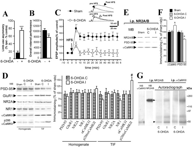
Figure 1.
Motor performances, synaptic plasticity, and alteration of αCaMKII-PSD-95 binding to the NMDA receptor complex in sham- and 6-OHDA-lesioned striatum. A, A limb-use asymmetry test was performed in sham-operated and 6-OHDA denervated rats (n = 8 in each group). 6-OHDA-lesioned rats preferentially use the limb ipsilateral to the lesion (***p < 0.0001; 6-OHDA vs sham). ipsi, Ipsilateral; contra, contralateral. B, Coordinated locomotor activity on a rotarod is significantly impaired after dopamine denervation (**p < 0.001; 6-OHDA vs sham). C, HFS of corticostriatal fibers induced LTP in sham-operated rats (filled circles; p < 0.01; EPSP amplitude after vs before HFS; n = 25) but not in 6-OHDA-lesioned animals (open circles; p > 0.05; EPSP amplitude after vs before HFS; n = 20). D, Effects of 6-OHDA lesioning on the NMDA receptor complex in the rat striatum. Striatal homogenates (left) and TIFs (right) from sham- and 6-OHDA-lesioned animals were analyzed by Western blot analysis with PSD-95, GluR1, NR2A, NR1,αCaMKII, and active p286-αCaMKII antibodies. The same amount of protein was loaded per lane (*p < 0.05; #p < 0.01; 6-OHDA I vs 6-OHDA C; n = 8 for each group). E, TIF proteins from sham- and 6-OHDA-lesioned animals were immunoprecipitated with an NR2A-NR2B polyclonal antibody. Western blot analysis was performed in the immunoprecipitated (i.p.) material with αCaMKII and PSD-95 antibodies. F, Quantitative analysis of Western blot performed on coimmunoprecipitated material. *p < 0.01 (6-OHDA I vs sham). G, TIF proteins were phosphorylated under conditions known to maximally activate CaMKII and then immunoprecipitated with anti-NR2A-NR2B (left) or anti-CaMKIIα (right). Autoradiography obtained after immunoprecipitation shows two major phosphorylated protein bands: a 50 kDa band (bottom arrow) pointing to autophosphorylated αCaMKII and a 170 kDa band (top arrow) indicating phosphorylated NR2A-NR2B. Left lanes, Identification by Western blot of NR2A-NR2B and αCaMKII in the immunocomplex. WB, Western blot; I, striatum ipsilateral to the lesion; C, striatum contralateral to the lesion.

