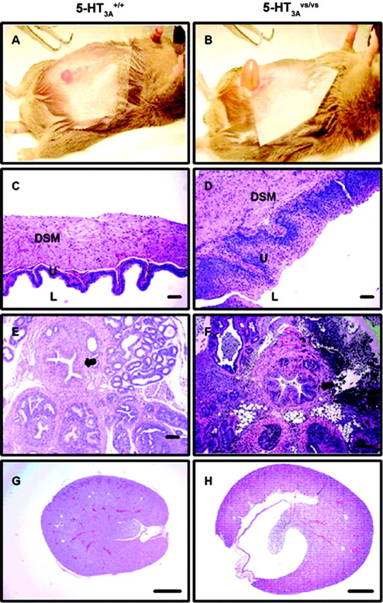
Figure 5.

A-H, Histopathology of the lower urinary tract from 5-HT3A+/+ (A, C, E, G) and 5-HT3Avs/vs (B, D, F, H) mice. A, B, Representative photograph of the hyperdistended urinary bladders seen in 5-HT3Avs/vs (B) compared with 5-HT3A+/+ (A) mice. C, D, H and E stained sections of the urinary bladder wall highlighting the lumen (L), urothelial mucosal layer (U), and the detrusor smooth muscle layer (DSM). E, F, H and E stained sections of the prostatic urethra and surrounding prostatic tissue. Note the increased thickness of the urinary bladder wall (D vs C) and prostatic urethra (arrow in F vs E) in 5-HT3Avs/vs compared with 5-HT3A+/+ mice, with associated mucosal hyperplasia and smooth muscle hypertrophy. In addition, extensive glandular and periglandular inflammatory cell infiltrate can be seen in the prostatic sections of male 5-HT3Avs/vsmice(F).G,H, Longitudinal sections of kidney from 5-HT3A+/+ (G) and5-HT3Avs/vs (H) mice showing the markedly distended renal pelvis in 5-HT3A mutant mice as a consequence of chronic lower urinary tract obstruction. Scale bars: C, D, 50 μm; E-H, 100 μm.
