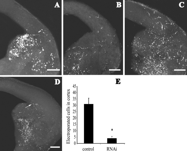
Figure 2.
Electroporation of lhx6 silencing constructs in E13.5 mouse brain slices. A-C, The GE was electroporated with control vector/pEGFP, and tangential migration was visualized after 24 hr (A), 48 hr (B), and 72 hr (C). More GFP cells were found in the cortex with time. Electroporation of siRNA vector/pEGFP resulted in labeled neurons dispersing within the GE but not entering the cortex (D). Sometimes, neurons (arrow) would accumulate at the corticostriatal boundary. Scale bars, 200 μm. E, Graph depicts quantification of cell migration from the GE electroporated with control vector (31 ± 4.7 cells within the cortex) and siRNA vector (4 ± 1.5 cells at corticostriatal boundary; Student's t test; p < 0.0001).

