
Figure 3.
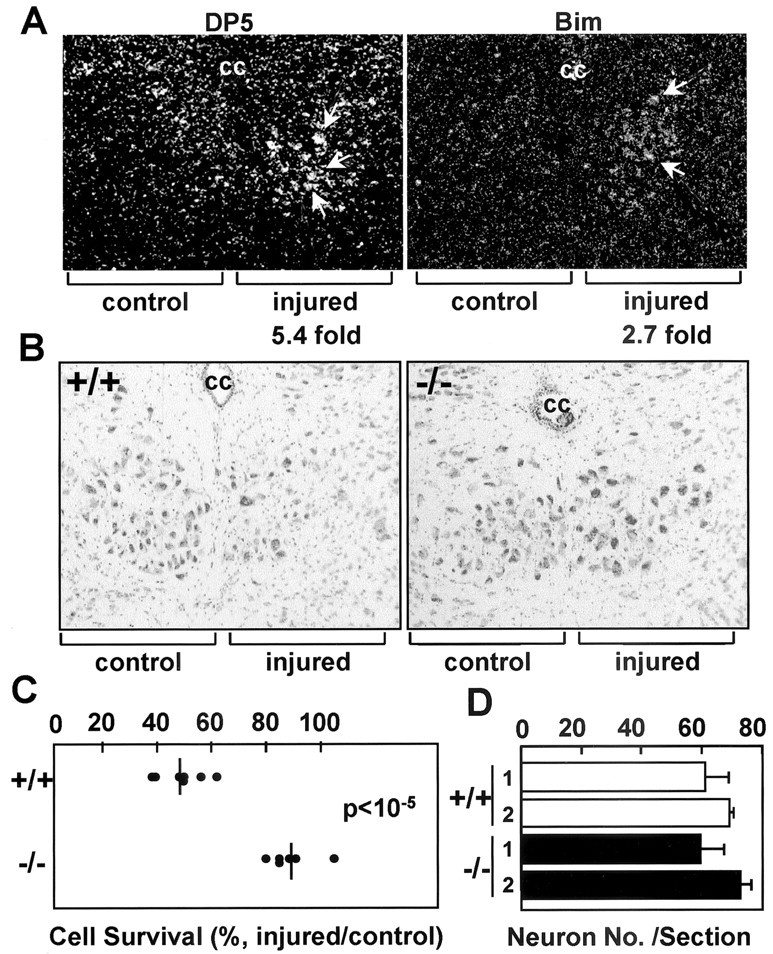
DP5 deletion protects motoneurons from axotomy-induced cell death. A, In situ hybridization of hypoglossal nuclei from 8-week-old mice with dp5- and bim-labeled probes 7 d after hypoglossal nerve transection. Injured and uninjured (control) sides are shown. The fold induction of dp5 and bim in injured neurons (compared with the uninjured side) calculated by image scanning is indicated. B, Coronal sections of the hypoglossal nuclei from DP5+/+ and DP5–/– mice 35 d after axotomy. The injured and control (contralateral) sides are shown. Motoneurons were stained with thionine. cc, Central canal. C, Survival of motoneurons from DP5+/+ and DP5–/– mice 35 d after hypoglossal nerve transection. The number of neurons in the injured and uninjured hypoglossal nuclei was determined by counting seven different sections per animal. Results represent the number of motoneurons in each axotomized nucleus as a percentage of the number in its contralateral (uninjured) nucleus. The DP5+/+ (n = 7) and DP5–/– (n = 6) mice were significantly different (p < 10–5; Student's t test). D, Number of neurons in hypoglossal nuclei (uninjured side) from 8-week-old DP5+/+ and DP5–/– mice. Values (number of neurons) represent the means ± SD derived from seven sections per animal. Results derived from two DP5+/+ and two DP5–/– mice are shown.