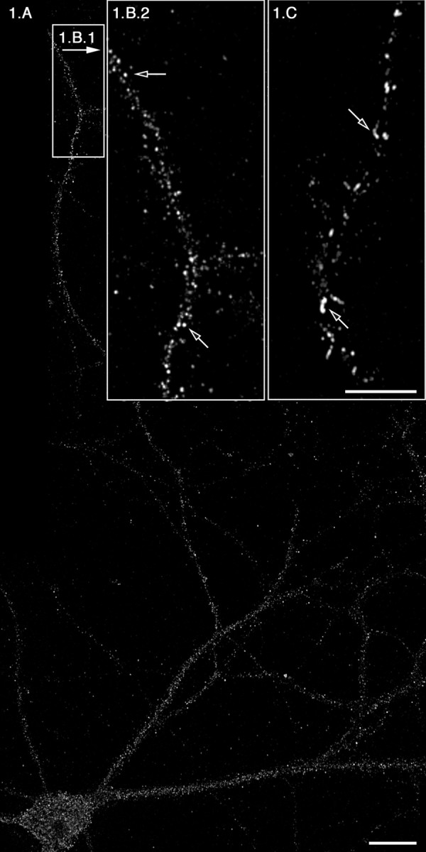
Figure 1.

ER export occurs efficiently and regularly at distances >350 μm from the soma. Rat hippocampal neurons were rinsed with PBS, permeabilized, and washed as described in Materials and Methods. Cells were then incubated in the presence of RLC and 5 μg of Sar1-GTP (A, B1, B2) or with Sar1-GTP only (C). At the end of the incubations, the distribution of Sec13 (A, B1, B2) and Sar1 (C) was determined using IF microscopy. The open arrows in B2 denote punctate VTCs. The open arrows in C denote ER membrane tubules coated with Sar1. B2 and C are higher magnifications of tertiary dendrites that are >350 μm away from soma. Scale bars: A, 20 μm; (in C) B2, C, 10 μm.
