Figure 1.
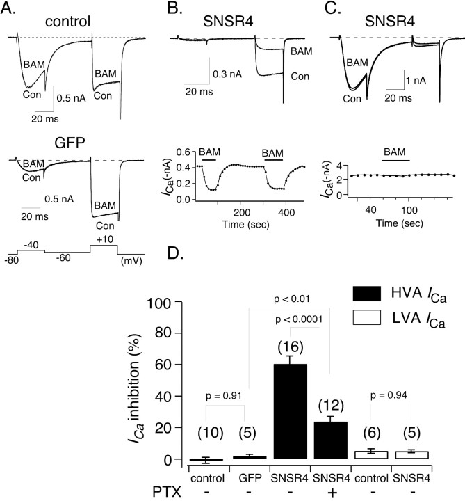
Heterologously expressed hSNSR4 couples to HVA but not LVA Ca2+ channels in rat DRG neurons. A, Representative whole-cell patch-clamp ICa traces acquired in the absence or presence of the peptide agonist BAM8-22 (BAM; 3 μm) from a control (Con; top) or an EGFP-expressing DRG neuron (middle). The voltage protocol used to evoke ICa for experiments described in Fig. 1 is illustrated at the bottom. The current evoked at -40 mV arises from LVA Ca2+ channels, whereas the current evoked at +10 mV (after an inactivating interpulse) arises primarily for HVA Ca2+ channels. B, Representative current traces (top) illustrating marked inhibition of ICa evoked (+10 mV) after application of BAM8-22 to a DRG neuron expressing hSNSR4. The neuron illustrated exhibited virtually no LVA Ca2+ channels. The time course of inhibition is shown at the bottom. C, Representative current traces (top) obtained in the absence or presence of BAM8-22 from an hSNSR4-expressing DRG neuron with a large component of LVA-type Ca2+ channels. The time course of inhibition is shown at the bottom. D, Summary bar graph comparing mean BAM8-22-induced inhibition of HVA (filled) and LVA (open) ICa under experimental conditions listed below each bar. HVA ICa amplitude was measured 10 msec from the start of the voltage step to +10 mV, whereas LVA ICa amplitude was measured 15 msec after initiation of the voltage step to -40 mV. Heterologous expression was accomplished using recombinant adenovirus.

