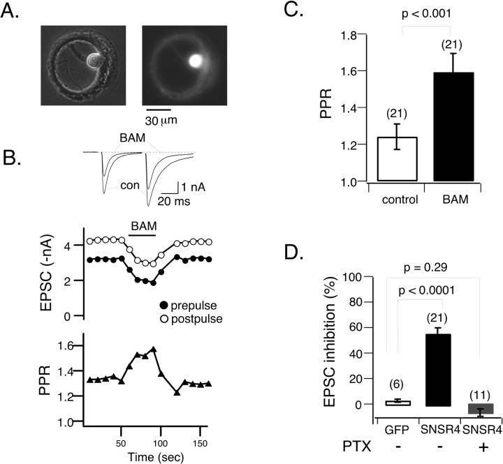
Figure 7.
Activation of hSNSR4 induces presynaptic inhibition in rat hippocampal neurons. A, Recombinant adenoviruses-mediated coexpression of hSNSR4 and EGFP in a hippocampal neuron cultured on a microisland of permissive substrate for 2 weeks. The phase-contrast (left) and fluorescent (right) micrographs were taken from the same cell. con, Control. B, Top, Representative whole-cell current traces obtained from a neuron expressing hSNSR4 showing depression of the EPSCs during application of BAM8-22 (BAM; 3 μm). EPSCs were evoked by pairs of 2 msec depolarizing commands (50 msec interval between prepulse and postpulse) applied at 0.1 Hz. B, Middle, Time course of BAM8-22-induced inhibition of EPSCs evoked by prepulse (filled circles) and postpulse (open circles) depolarizations. B, Bottom, PPR increased during application of BAM8-22, indicating presynaptic locus of inhibition. C, Bar graph depicting mean (±SEM) PPR in the absence (open) or presence (filled) of BAM8-22 in neurons expressing hSNSR4. D, Bar graph comparing BAM8-22-induced mean (±SEM) percentage inhibition of EPSCs (evoked by prepulse) in neurons expressing EGFP (open, left bar) or hSNSR4 either untreated (filled, middle bar) or after overnight treatment with PTX (filled, right bar). The number of neurons tested is indicated in parentheses.

