
Figure 2.
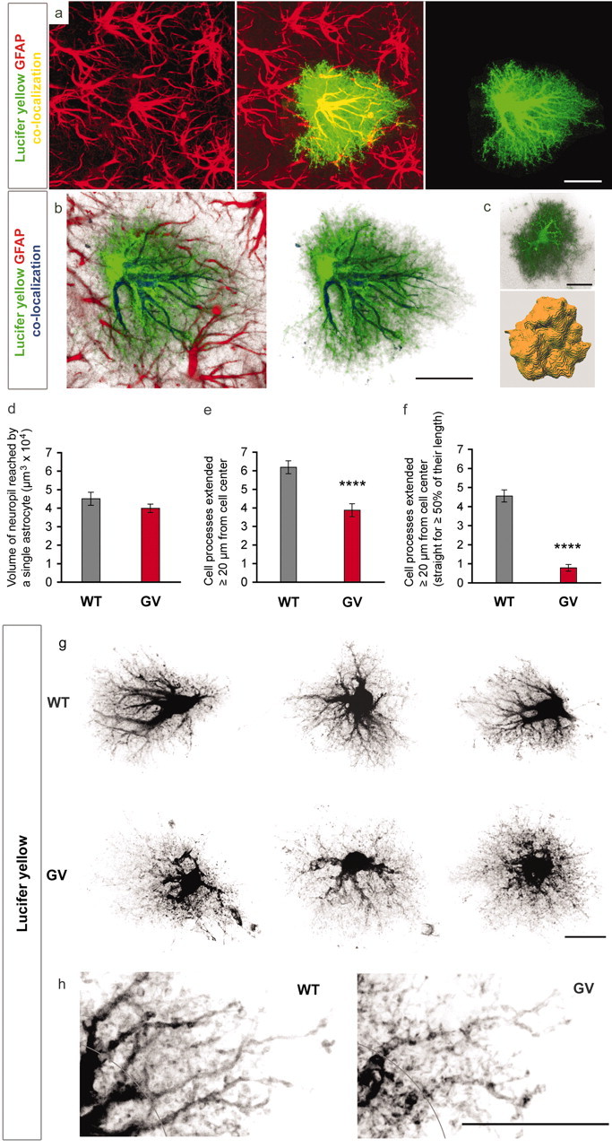
Dye filling reveals an altered morphology of astrocytic processes in GFAP-/-Vim-/- (GV) reactive astrocytes. a, Filling of individual reactive astrocytes with lucifer yellow shows many fine cellular processes that are not visualized by GFAP antibodies. a, b, GFAP-containing intermediate filaments are abundant in the main astrocytic processes but not detectable in many fine and terminal processes. Green, Lucifer yellow; red, GFAP. c, d, Three-dimensional reconstruction (c) showed that GFAP-/-Vim-/- and wild-type (WT) reactive astrocytes reach comparable volume of brain tissue at 4 d after entorhinal cortex lesion (d), but their processes are less hypertrophic; GFAP-/-Vim-/- astrocytes compared with wild-type have fewer long processes (e, ****p < 0.0001; g, h) as well as fewer straight processes (f, ****p < 0.0001; g, h). Black, Lucifer yellow. A distance of 20 μm from the center of the soma is indicated in h. Scale bars, 20 μm.