
Figure 6.
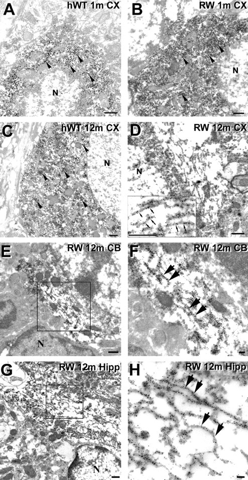
Intraneuronal tau inclusions in the RW tau Tg mice are composed of straight filaments. A-H, Preembedding immuno-EM using 17026 antibodies show tau immunostaining in neuronal perikarya of the hWT and RW tau Tg mice. Neocortical neurons of a 1-month-old hWT mouse (A), a 1-month-old RW mouse (B), and a 12-month-old hWT mouse (C) demonstrate diffuse tau staining (arrowheads) but no obvious tau filaments. D, Randomly oriented tau-positive straight filaments are demonstrated in the perikaryon of a neocortical neuron of a 12-month-old RW tau Tg mouse, and a higher-power view of these filaments (small arrows) is shown in the inset. E-H, Preembedding immuno-EM using the 17026 antibody shows tau-immunoreactive straight filaments in neurons from several CNS areas of a 12-month-old RW mouse. Higher-power views of the tau filaments (large arrows in F, H) are from the boxed areas in the adjacent panels showing lower-power views (E, G). The tau-immunoreactive filaments were found in the cerebellum (E, F) and hippocampus (G, H). N, Nucleus. Scale bars: A-E, G, 500 nm; F, H, 100 nm.