Figure 1.

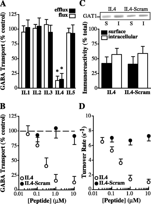
GAT1 IL4 domain inhibits GABA transport rates. A, IL4 fusion protein inhibits GABA flux and efflux. Oocytes expressing GAT1 were injected 15 min before assay with fusion proteins corresponding to each of five GAT1 intracellular domains (IL1-IL5; ∼1 μm final concentration) and were subjected to [3H]GABA flux (open bars) and efflux (filled bars) assays. Data are plotted relative to nonpeptide-injected oocytes and are from three experiments (4-7 oocytes per condition per experiment). B, Synthetic IL4 peptide inhibition of GABA uptake is concentration dependent. GAT1-expressing oocytes were acutely injected with various concentrations of IL4 peptide (open circles) or scrambled IL4 peptide (filled circles) and subjected to [3H]GABA flux assays. Data are plotted relative to nonpeptide-injected oocytes and are from three oocyte batches (5-9 oocytes per data point). C, IL4 peptide does not alter GAT1 surface expression. GAT1-expressing oocytes were acutely injected with 1 μm final concentration of IL4 or scrambled IL4 peptide and subjected to biotinylation. Immunoblot shows GAT1 immunoreactivity for avidin-bound (S, surface; filled bars) and nonbound (I, intracellular; open bars) fractions. Data from two experiments (20 oocytes per condition per experiment) are quantified relative to total GAT1 expression. D, IL4 peptide reduces GAT1 turnover rate in a concentration-dependent manner. Oocytes were treated as in B and evaluated using two-electrode voltage clamp. Data are from 6-13 oocytes per data point.
