
Figure 5.
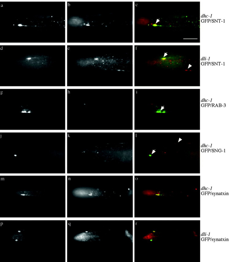
Localization of synaptic proteins in dynein mutants. In all images, arrows point to misaccumulations at the tip of ALM neurons. Arrowheads point to synaptic vesicle protein in an SAB neuron. a-f, Double labeling of GFP (green, first image of set) and SNT-1 (red, second image of set) immunoreactivity. a-c, dhc-1(js121); jsIs37. d-f, dli-1(js351); jsIs37. SNB-1::GFP misaccumulations in both genotypes contain SNT-1 immunoreactivity. g-i, Double labeling of GFP (green, first image of set) and RAB-3 (red, second image of set) immunoreactivity of dhc-1(js121); jsIs37. j-l, GFP (green, first image of set) and transmembrane protein SNG-1 (red, second image of set) immunoreactivity of dhc-1(js121); jsIs37. SNB-1::GFP misaccumulations do not contain either RAB-3 or SNG-1 immunoreactivity. m-r, Double labeling for GFP (green, first image of set) and UNC-64 syntaxin (red, second image of set) immunoreactivity. m-o, dhc-1(js121); jsIs37 SNB-1::GFP containing misaccumulations also contain syntaxin. p-r, dli-1(js351); jsIs37. Two SNB-1::GFP misaccumulations are present in the nose, one of which contains syntaxin (arrow) and one of which does not (arrowhead). In general, ∼40-50% of SNB-1::GFP misaccumulations contain syntaxin. The buccal cavity of the animal often shows nonspecific reactivity with many antibodies; this is seen in b, c, e, f, i, n, o, q, and r. Scale bar, 10 μm.