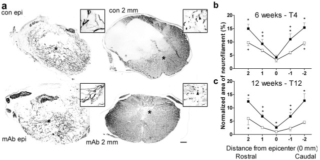
Figure 6.
Anti-CD11d mAb treatment increases neurofilament in the injured cord after SCI. Neurofilament-stained sections taken at 6 weeks after T4 SCI are illustrated at low and higher (inset) power at the lesion epicenter and at 2.0 mm caudal to the epicenter in control and mAb-treated rats (a). These sections are within 40 μm of those stained for myelin illustrated in Figure 5a. Within the lesion, neurofilament fibers were mostly arrayed in disorganized bundles in control and mAb-treated rats. At 2.0 mm caudal to the lesion, the neurofilament was more intact throughout the gray and white matter, and the greater integrity of the neuropil after anti-CD11d mAb treatment is again reflected by the smaller cavitation in the section. White matter axon bundles were aligned more normally at 2 mm from the lesion site, and greater numbers were visualized in cross-section in the treated rats (arrowheads in insets). *Indicates area from which high-power inset was taken. Scale bar is 200 μm for the low-power photomicrographs and 10 μm for the insets. Treatment effects on normalized areas of neurofilament are illustrated after SCI at T4 (b,6 weeks) and T12 (c,12 weeks). □, Control rats (b, c, n = 5). ▪, mAb-treated rats (b, n = 7; c, n = 6). *p < 0.05 compared with control rats. +Shortest distance where area is larger than area at epicenter, p < 0.05.

