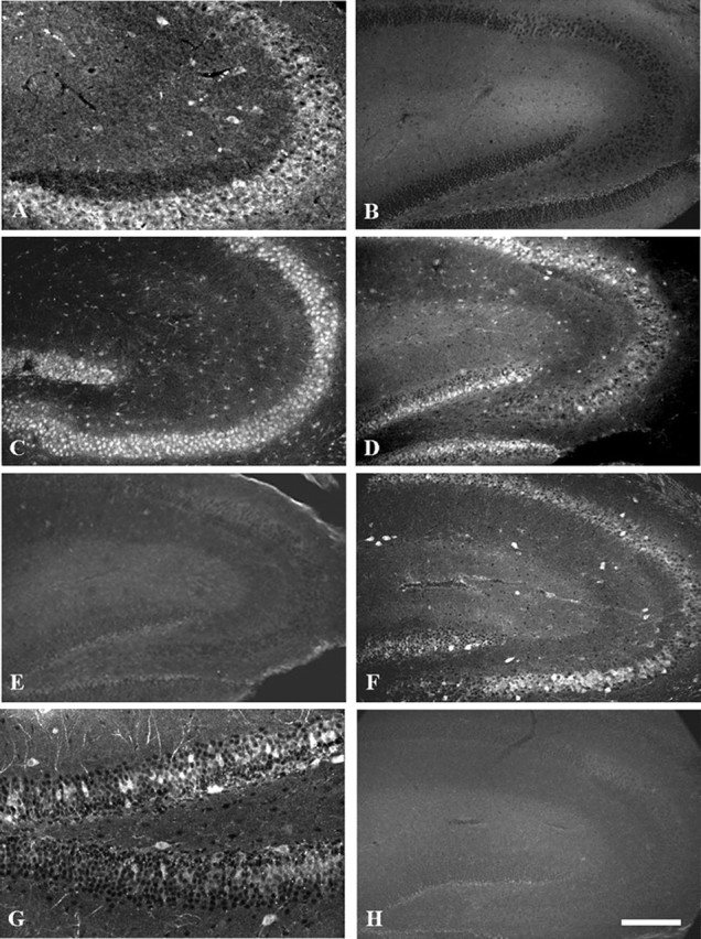
Figure 10.

β1 (-/-) mice show abnormal expression of Nav 1.1 and Nav 1.3 in the hippocampus. A, β1 (+/+) mice stained with anti-β1ex. B, β1 (-/-) mice illustrating the lack of anti-β1ex staining and the specificity of the null mutation. C, β1 (+/+) mice stained with anti-Nav1.1. D,β1(-/-) mice stained with anti-Nav1.1 illustrating lack of staining of many neurons in the CA2/CA3 region and in the outer leaflet of the dentate gyrus compared with controls (C). E, β1 (+/+) mice stained with anti-Nav1.3. F, β1 (-/-) mice stained with anti-Nav1.3 illustrating increased staining in interneurons of the hippocampus. G, Higher magnification of the dentate gyrus labeled with anti-Nav 1.3 antibodies illustrating increased staining in neurons located in this region of β1 (-/-) mice. H, Tissue section incubated with no primary antibodies to illustrate the specificity of antibody staining. Scale bars, 200 μm.
