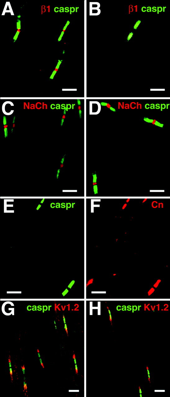
Figure 4.

Sciatic nerve nodes of Ranvier in β1 (-/-) mice. Sciatic nerves were isolated from β1 (+/+) and β1 (-/-) P17-P19 mice and prepared for immunocytochemistry, as described in Materials and Methods. Immunofluorescent staining obtained from antibodies directed against pan-sodium channels, caspr 1, contactin, and Kv1.2 appeared to be unchanged in the mutant mice compared with control. A, β1 (+/+) mice: β1 subunits (red), caspr 1 (green). B, β1 (-/-) mice: β1 subunits (red), caspr 1 (green). There is no staining for β1 subunits. C,β1(+/+) mice: pan-sodium channels (red), caspr 1 (green). D,β1(-/-) mice: pan-sodium channels (red), caspr 1 (green). E,β1(-/-) mice: caspr 1 (green). F,β1(-/-) mice (identical section as in E): contactin (red). G, β1 (+/+) mice: caspr 1 (green) and Kv1.2 (red). H, β1 (-/-) mice: caspr 1 (green) and Kv1.2 (red). Scale bars, 10 μm.
