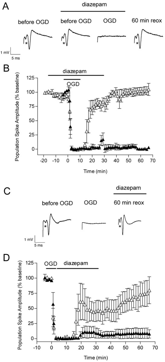
Figure 10.

Effect of OGD and diazepam (applied before and after OGD) on the evoked population spike in area CA1 pyramidal cells. A, Diazepam (5 μm) was added 15 min before OGD, and it was removed from the slice chamber 30 min after OGD. Representative traces are shown before, during, and 60 min after OGD in the presence of diazepam. The first trace was recorded before the addition of diazepam. B, The time course is shown for changes in the population spike amplitude recorded from pyramidal CA1 neurons in the absence (▴) and in the presence (▵) of diazepam in control and OGD slices. Note the small decrease in population spike amplitude (p < 0.05) starting at –12 min because of the presence of diazepam. To reduce crowding of data points, each data point is the average of three consecutive recordings that were made every 30 sec. Data are the means ± SEM of four slices per condition. C, Diazepam (5 μm) was added immediately after OGD, and it was removed from the slice chamber 30 min later. Individual recordings are shown for a control hippocampal slice before, during, and 60 min after 7 min OGD plus diazepam. D, The time course is shown for changes in the population spike amplitude recorded from pyramidal CA1 neurons in the absence (▴) and in the presence (▵) of diazepam in control and OGD slices. Each data point is the average of three consecutive recordings that were made every 30 sec. Data are the means ± SEM of five slices per condition. The recovery of the population spike amplitude by 60 min of reoxygenation was not different from baseline (p > 0.05; ANOVA and Tukey's multiple comparison test).
