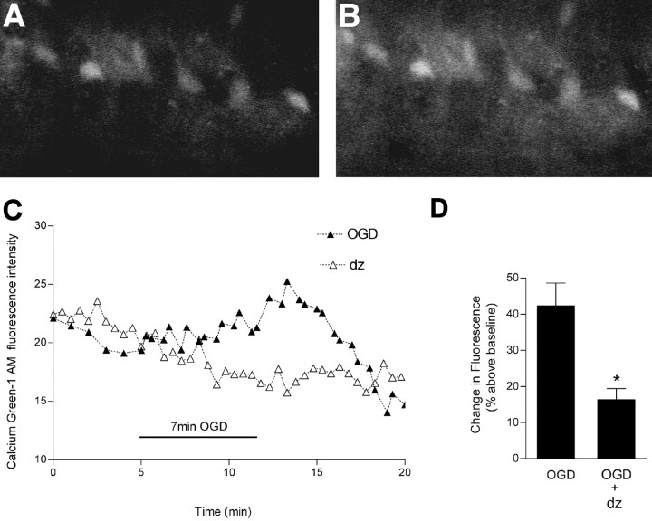
Figure 9.
Effect of diazepam on OGD-induced changes in intracellular Ca2+. Slices were loaded with Calcium Green-1 AM and subjected to OGD for 7 min. A, Video image of area CA1 pyramidal cells shows Calcium Green-1 fluorescence during baseline recording. B, An increase in fluorescence is indicated after OGD. C, Continuous recording of Calcium Green-1 fluorescence before, during, and after OGD in representative slices. In these experiments, diazepam (5 μm) was added to the superfusion buffer at the onset of OGD to affect the immediate rise in intracellular Ca2+. D, The peak increase in intracellular Ca2+ occurred ∼10 min after the onset of OGD. Values are expressed as the percentage increase in fluorescence intensity [(ΔF/F) × 100] of Calcium Green-1 above baseline. Data are the means ± SEM of six and five cells per condition, respectively. *p < 0.01 versus OGD; unpaired Student's t test.

